November 03, 2026
Gouri Nayathil and Amy Rivers spent eight weeks investigating pulmonary fibrosis, supported by Action for Pulmonary Fibrosis and the British Association for Lung Research (BALR).
By supporting opportunities like these, we’re helping to inspire the next generation of respiratory researchers and encouraging more early-career scientists to pursue careers investigating lung diseases.
Bettina Schock, BALR Partnership Secretary, said:
“BALR’s goal is to support the education and career development of lung researchers, thereby strengthening pulmonary research in the UK. Through the continuing support of our summer studentship scheme, APF enables us to fulfil this goal and gives students an important career directing opportunity.
“We vastly appreciate this important collaboration, which will increase the number of highly motivated respiratory researchers to improve the lives of people with pulmonary fibrosis.”
Gouri’s research project
Certain genetic variants may increase an individual’s risk of developing respiratory diseases including idiopathic pulmonary fibrosis (IPF), asthma and COPD. However, it is not fully understood how these genetic variants contribute to disease. Understanding this could help researchers uncover the biological processes driving disease and identify new targets for treatment.
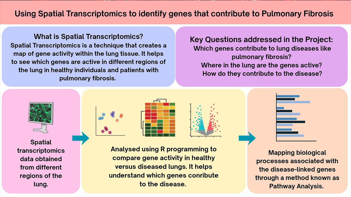
Under the guidance of Dr Robert Hall at the University of Nottingham, Gouri analysed existing data from different parts of human lung tissue to look at how these disease-linked genes behaved in IPF and COPD lung tissue and explored the biological processes underlying their association with disease.
She used techniques including spatial transcriptomics, which can show where genes are active within lung tissue, alongside clinical data. By identifying and ranking the genes most closely linked to disease-related changes, the project aimed to help researchers prioritise promising targets for future treatments.
Gouri said: “I applied for the studentship as I wanted to contribute to research that translates into improved patient outcomes. With a career aspiration in translational research, I was keen to work on respiratory diseases such as IPF and COPD, which affect millions of people globally.
“Throughout the studentship, I enjoyed working with real-world datasets and was able to develop my computational skills while learning how to troubleshoot problems, interpret results and think critically about research findings.
“Comparing gene expression between diseased and healthy samples helped identify genes and biological mechanisms that may contribute to these diseases. These findings could be particularly helpful in improving future treatments for patients.”
Amy’s research project
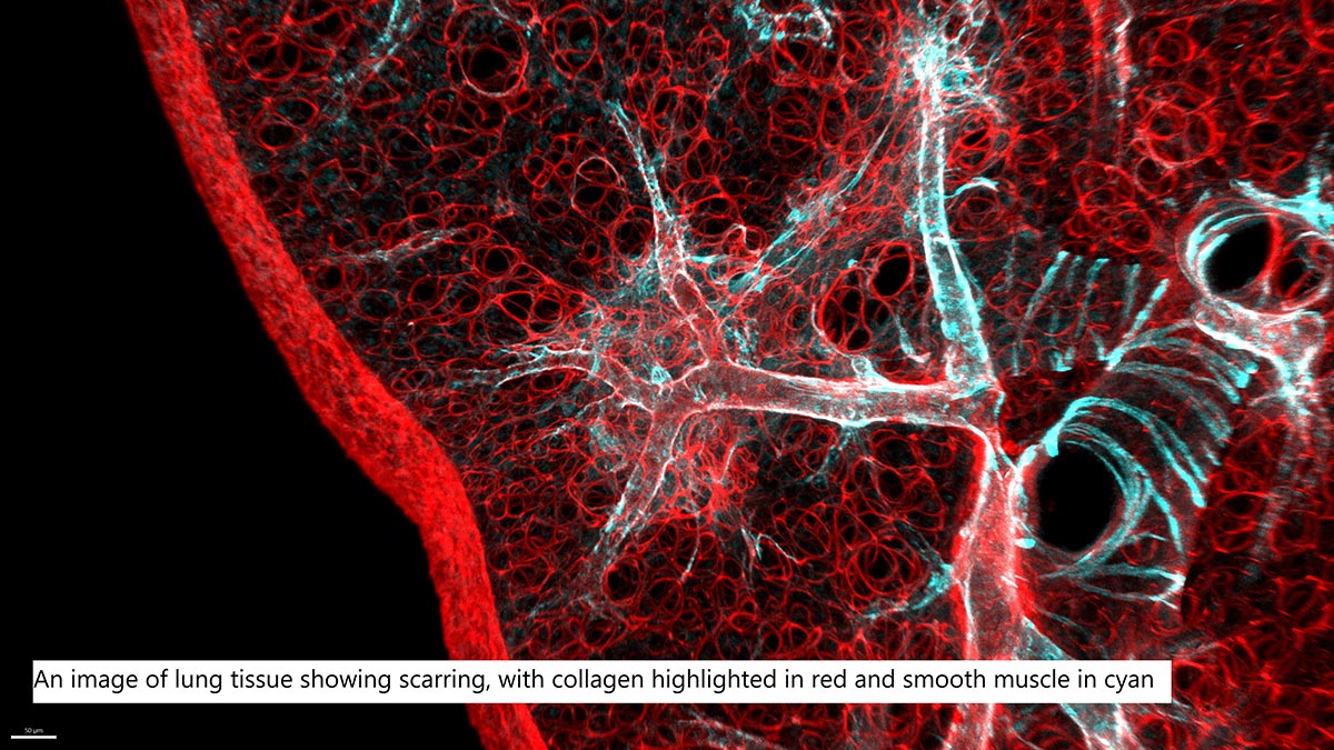
Pulmonary fibrosis causes scar tissue to build up in the lungs, making them stiffer and harder to expand. Detecting this scarring can be difficult because doctors sometimes examine very thin slices from lung biopsy samples. If fibrosis is patchy or at an early stage, some signs may be missed.
Under the guidance of Dr Simon Cleary at King's College London, Amy tested a new way of looking at lung tissue in three dimensions. The technique used fluorescent dyes to highlight collagen, a key protein that forms fibrotic scar tissue, alongside ‘tissue clearing’ methods that make samples transparent so they can be imaged without being cut into thin slices.
She tested the approach on mouse and human lung samples to see how well it could detect early signs of fibrosis. The project aimed to explore whether this method could eventually help improve detection of pulmonary fibrosis and support research into new treatments.
Amy said: “I was delighted to receive a BALR Summer Studentship with Action for Pulmonary Fibrosis, working with Dr Simon Cleary on 3D imaging approaches for studying pulmonary fibrosis.
“The project gave me training in both standard and novel microscopy techniques, as well as manual and automated image analysis. It strengthened my laboratory and scientific communication skills, and I learned how to develop and test a new method.
“We found that fast green FCF, a small molecule that binds fibrillar collagen, allows highly sensitive and specific fluorescence imaging of fibrosis in mouse and human lung samples. We worked on approaches to use this imaging method with precision-cut lung slices and whole biopsies, and it may be useful for improving how we measure fibrosis in preclinical studies and for early detection of fibrotic changes in clinical pathology.”